The IBC Network Foundation founder Terry Arnold has chronicled her journey with lymphedema through social media posts. Here we have pulled them all together in one article for you to follow. This post will continue to be updated.
We also wanted to share a link to a great article from Fred Hutch Cancer Center that will answer some of your lymphedema questions. Terry has had all of the surgeries mentioned. Read the entire article here.
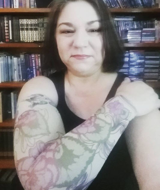
“I’ve taken a series of photos so you can see my arm looks like after using my new compression pump. The first picture shows my arm before the compression pump was used. The other pictures show my arm after the 50-minute session, and you can see there’s deep intentions into my arm. My arm is literally deflated. The fluid is being moved from my arm, and I literally pee the fluid out of my body. This has been working so well. I’ve actually lost about three pounds in the last month. That fluid has got to come out somehow. And if you’re not urinating a lot after using compression, you’re not getting the effect of the compression. The fourth picture has a mark. Because I want you to notice that there’s no indentions from mid forearm towards my wrist. There is a reason for that. That is the area that has the least channels in my body according to the testing that we have done. The compression sleeve goes all the way down to the tips of my fingers. So hopefully soon I will start seeing indentions in that problem area because I know the channels are growing back. I’ve seen evidence in our testing. If you’ve seen previous posts where I show the time tracking through my body…. You’ll know what I’m talking about.”


The new compression jacket has arrived! It’s a jacket that goes down to my waist and has one arm sleeve.


Researcher Melissa Alrich, PhD is explaining to me the actual make up of a lymphatic vessel. It is a thin tube that carries lymp (lmphatic fluid) and white blood cells through the lymphatic system. What she continues to explain is that the system does not have a pump, like the heart to move blood through the body, but depends on body movement and pressure. On the outer wall of the tube is a think muscle. This muscle, just for a simple explanation sake is not a muscle that gives pressure, (more like a barrier wall). However this muscle is damaged by chemotherapy, and that damage allows the fluid to “weep” out of the tube, and become leaky and more porous. This is part of the reason even with good care and massage, the effected limb can still swell. There is not a way to get the “weepy” fluid back in to the tube. You can see in later pictures a clean line of a tube, with good flow but also places where there is a “cloud” of green fluid. The green is the dye from the testing to help make it easier to see the movement. A cloud of green, is where the fluid has wept out, and backed up into the limb.

Close up of graphic showing the difference of a tube that is carrying fluid properly and also an example of where the fluid can weep out of the tube, collecting in the effected limb.

This is a close up of my hand. The dye to trace the flow of fluid is shot between the fingers. Personally I did not find it hurt, as they do prepare with numbing cream. Some do say the dye can sting but my hand from such advanced LE, has a great deal of numbness, so I didn’t feel it.

This is a shot of a video that was recorded in the testing.


